Other
|
Multiscale analysis of bone qualitative changes in human subchondral bone as a basis for the pathogenesis of osteoarthritis
Funding line:
First and Second Applications

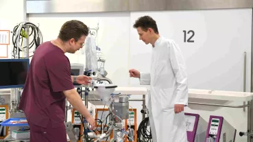
Dr. Julian Stürznickel (left), Dr. Felix N. Schmidt (front-right) and PD Dr. Dr. Tim Rolvien (back-right) analyzing the subchondral bone in the osteoarthritic femoral head using high-resolution computed tomography (HR-pQCT).
(© University Medical Center Hamburg-Eppendorf, Tim Rolvien)
While degeneration of the articular cartilage is the main feature of osteoarthritis (OA), complex mechanisms influence its development and progression. In this regard, a special role is attributed to the bone below the cartilage (so-called subchondral bone). We are performing a comprehensive imaging and biomechanical analysis of the subchondral bone of human samples with different grades of OA severity. The aim of the project is to decipher the cellular, bone quality and biochemical changes with identification of stage-specific and topographic patterns. This way, the role of subchondral bone in OA development will be elucidated, which may help in the search for new therapeutic options.
Here you can find further information.