Innovative PET tracers for non-invasive molecular characterization of brain tumors and evaluation of theranostic options

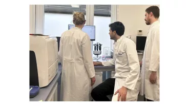
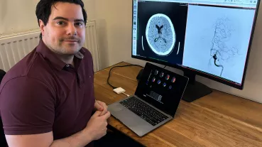
Gliomas are primary brain tumors and still associated with an extremely poor prognosis, despite ongoing research and development of therapeutic approaches. In the standard imaging of brain tumors by means of magnetic resonance imaging (MRI), the depiction of the tumor extent is highly challenging, so that the treatment planning for surgical removal and irradiation of the entire tumor is correspondingly difficult. Positron emission tomography (PET) using radioactively labeled substances can be used to obtain valuable additional information about the extent and vitality of gliomas.
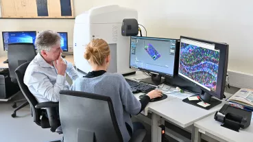
The aim of this research project is to improve brain tumor imaging by combining MRI and PET imaging using novel radiotracers. In particular, a prospective study examines whether the tumor borders can be delineated better and whether more aggressive tumor parts within a glioma can be reliably identified in order to enable tailor-made therapy for the individual patient and to improve the therapeutic outcome. In a further part of the research project, novel nuclear medical therapeutic approaches will be evaluated in which tumor cells are destroyed by radioactive radiation.
Overall, an improved delineation and characterization of brain tumors as well as potential innovative therapies should optimize the treatment of glioma patients and sustainably improve their survival.
Here you can get further information.